3-D Mesoscale Soft X-ray microtomography for low contrast biological or soft samples
Reconstructing the 3-D structure of small objects
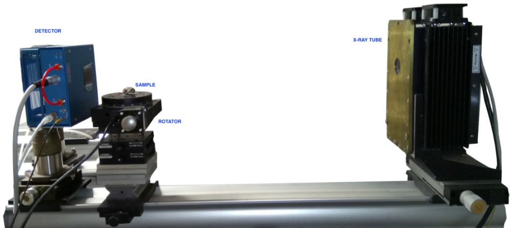
Experimental set-up for micro-tomographic analyses
A fusion-based mesoscale soft X-ray microtomograph with energy discrimination and in energy bands was developed and assembled at the ENEA Laboratories in Frascati (Italy) for reconstructing the 3-D structure of small objects (a few cubic centimeters), with moderate spatial resolution (a few microns) but for samples pretty transparent to X-rays and with very low contrast, like biological samples or light (soft) materials (see fig.1).
The technology of soft X-ray imaging with energy discrimination and in energy bands has been developed in many years for diagnosing magnetic fusion plasmas, produced in machine called tokamak. In this domain, energy resolution is crucial to recognize X-ray emissions produced by impurities and fuel in the plasma. Impurities indeed emit at different energies and in different regions of the plasma. This novel approach was studied over many tokamaks all over the world (FTU, NSTX, Tore Supra, KSTAR, EAST, etc.).
Mesoscale X-ray microtomography applied to samples transparent to X-rays and with very low contrast
These competencies, developed in the contest of the european magnetic fusion program, were used for the mesoscale X-ray microtomography with energy discrimination for reconstructing the 3-D structure of small objects (a few cubic centimeters), with moderate spatial resolution (a few microns) but for samples pretty transparent to X-rays and with very low contrast, like biological samples or light (soft) materials The novel technique stems from the selection of the spectrum of the X-ray tube, chosen suitably depending on the sample, and the X-ray detection in this optimized energy band.
The non-fusion application of this system was developed by NIXT for the ENEA division Biodiversity and Ecosystem Services Laboratory, Santa Teresa Research Centre, La Spezia. The research project had the aim to design and develop 3D artificial reefs (‘mimics’) simulating Ellisolandia elongata biogenic coralline red algae reefs and studying the suitability of the reef ‘mimics’ as habitat providers and biodiversity facilitators in natural environments.
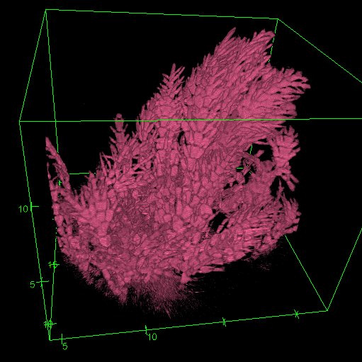
Microtomography was used to reproduce 3D images of natural Elissolandia elongata reef (fig.2), from which a design for algal fronds for the artificial reefs was created. NIXT laboratories provided the digital 3-D recontructions, for the different samples received, to the group in La Spezia and they developed the software to make a quantitative analysis. Performances of NIXT microtomograph were discussed with the group in La Spezia. The setup and optimization of microtomography was realised by NIXT Laboratory.
Beyond comercial tomographies with fusion tech
Biological samples such as algae cannot be investigated with commercial microtomographs, because they work with high energy X-rays and not efficient detection. Crucial for this technique is the 2-D imaging X-ray detector working in photon counting with energy discrimination; it is made of a CdTe semiconductor C-MOS imager with a matrix of 512 x 512 pixel, 55 um size. This mesoscale microtomography with energy control allowed the reconstruction of Ellisolandia algae, extremely important to study quality and health of marine environment otherwise not possible with commercial tomographies.